Are Dental X-Rays Safe? What Every Patient Should Know
Dental X-ray doses are among the lowest in medicine. Learn the four types dentists use, how often you need them, and the safety measures that protect you.
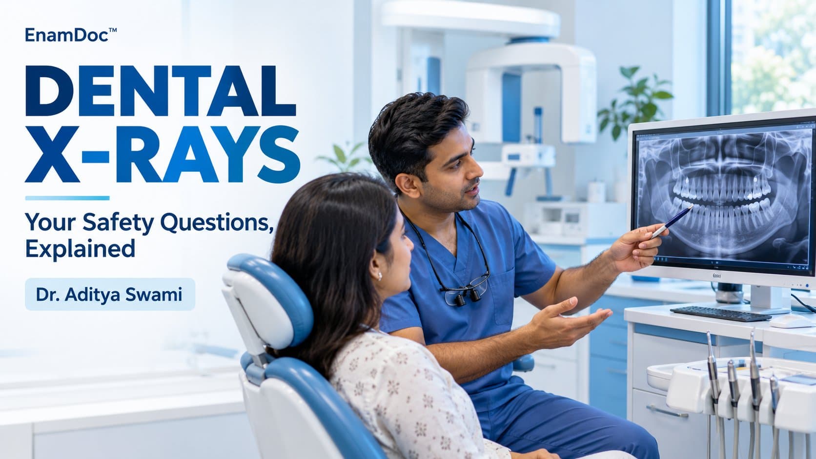
If you have ever hesitated before agreeing to dental X-rays, you are not alone. Radiation sounds alarming. But the radiation dose from a set of dental X-rays is lower than what you absorb during a short walk outdoors. Here is what the evidence actually says — and what your dentist is looking for when they request them.
Types of Dental X-Rays and What They Show
Bitewing X-Rays
The most common type, taken from the side with the film between your teeth. They reveal decay between teeth and changes in bone density caused by gum disease. Most adults have bitewings taken once a year or every 18 months.
Periapical X-Rays
Show the entire tooth from crown to root tip. Dentists use these to investigate pain, abscesses, or bone loss around a specific tooth.
Panoramic (OPG) X-Rays
A single rotating image that captures all your teeth, jaws, sinuses, and temporomandibular joints at once. Useful for assessing wisdom teeth, orthodontic planning, and jaw conditions.
Cone Beam CT (CBCT)
A 3D scan used for implant planning, root canal assessment, or complex orthodontic cases. It carries a higher dose than standard films, so it is reserved for specific clinical needs.
How Much Radiation Is Actually Involved?
Radiation exposure is measured in microsieverts (μSv). To give you context:
- A full-mouth series of 18 periapical films: approximately 35 μSv
- Four bitewing X-rays: approximately 5 μSv
- A panoramic X-ray: approximately 14–24 μSv
- A transatlantic flight: approximately 50–80 μSv
- Daily background radiation (natural sources): approximately 8 μSv per day
Dental X-rays represent a small fraction of the radiation you receive from natural sources every year. The risk is not zero — no radiation exposure is — but it is proportionate to the clinical benefit of finding problems early.
Who Should Have Them and How Often?
There is no single protocol that fits every patient. The frequency depends on your individual risk factors:
- Low risk (no recent decay, excellent oral hygiene): Bitewings every 18–24 months
- Moderate risk (previous decay, dry mouth, orthodontic appliances): Bitewings every 12 months
- High risk (active decay, heavy plaque, radiation therapy to the head): Bitewings every 6 months
Children typically need X-rays more often than adults because their teeth and jaws are developing rapidly and decay can progress faster.
Safety Measures Dentists Use
Lead Aprons and Thyroid Collars
Most dental clinics drape a lead apron over your chest, abdomen, and lap before taking X-rays. A thyroid collar specifically shields the thyroid gland, which is more sensitive to radiation. If your clinic offers one, always accept it — particularly for panoramic X-rays.
Rectangular Collimation
Older X-ray machines used a circular beam that exposed more tissue than necessary. Rectangular collimation limits the beam to roughly the size of the film, reducing scatter radiation by up to 60 percent.
Digital Sensors Instead of Film
Digital X-ray sensors require far less radiation than traditional film — typically 50–80 percent less. Most modern dental clinics in India have switched to digital systems.
Special Situations: Pregnancy and Children
Dental X-rays are generally considered safe during pregnancy when necessary. The dose to the developing baby from a lead-apron-protected dental X-ray is negligible. Emergency dental care during pregnancy should not be delayed because of radiation concerns. That said, routine surveillance X-rays are often deferred until after delivery if clinically appropriate.
For children, the same risk-benefit logic applies. Small mouths benefit most from early decay detection, and modern paediatric sensors minimise dose. If your child's dentist recommends X-rays, they have weighed the clinical indication against the dose.
When to Question an X-Ray Recommendation
X-rays should follow clinical need, not a fixed calendar. It is always appropriate to ask your dentist:
- Why are you recommending this X-ray today?
- Will the result change my treatment plan?
- Are previous X-rays from another clinic transferable?
A dentist who cannot answer these questions or who routinely takes X-rays without clinical justification is worth questioning. However, a recommendation backed by a sound clinical reason is in your interest — catching a cavity between teeth before it reaches the nerve is always preferable to a root canal.
Frequently asked
Frequently asked questions
How often should I have dental X-rays?
The frequency depends on your risk level. Low-risk adults may need bitewing X-rays every 18–24 months; those with a history of decay every 12 months; high-risk patients every six months. Your dentist tailors the schedule to your individual needs.
Are dental X-rays safe during pregnancy?
Yes, when clinically necessary. The radiation dose from a lead-apron-protected dental X-ray is negligible to the developing baby. Emergency dental care should not be delayed because of X-ray concerns, though routine surveillance films are often deferred until after delivery.
What is the difference between a bitewing and a panoramic X-ray?
Bitewing X-rays show the crowns and contact points of a section of teeth — ideal for catching interproximal decay. A panoramic (OPG) X-ray captures all teeth and both jaws in a single image, used for assessing wisdom teeth, jaw pathology, and orthodontic planning.
- dentalx-raysradiographradiationsafetybitewingOPGpanoramicx-ray
Keep reading