Dental X-Rays: Are They Safe, How Often Do You Need Them, and What Do They Show?
Patients often wonder whether dental X-rays are really necessary and whether the radiation is a concern. Here is what dental X-rays actually reveal, how often you genuinely need them, and the real answer on radiation safety.
Medically reviewed by Dr. Khanak Jain, BDS
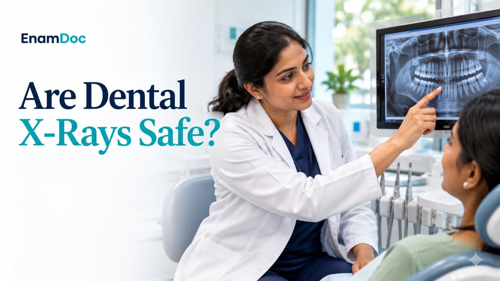
Dental X-rays are one of the most important diagnostic tools a dentist has — and one of the most questioned by patients. The concern about radiation is understandable and worth addressing directly. The short answer is that modern dental X-rays are extremely safe, and the far greater risk is the undetected decay, bone loss, or infection that X-rays would have caught. This article explains what different types of dental X-rays show, how often you actually need them, and exactly how much radiation is involved.
What Dental X-Rays Reveal That Clinical Examination Cannot
Even the most thorough visual and tactile examination misses things that X-rays reveal routinely:
- Interproximal decay: Cavities forming between teeth — completely invisible to the naked eye until they are large enough to affect the visible tooth surface. By the time interproximal decay can be seen, it is often already into the dentine.
- Bone levels: Gum disease causes progressive bone loss around the roots of teeth. X-rays show bone height, allowing the dentist to assess the severity of periodontal disease and track whether it is progressing or stable.
- Root conditions: Abscesses, cysts, and infections at the root tip (periapical pathology) are invisible clinically but clearly visible on X-rays. Some of these infections cause no pain until they are significant.
- Impacted teeth: Wisdom teeth and other teeth failing to erupt properly need X-ray assessment to determine their position relative to nerves and adjacent teeth — essential before any extraction.
- Existing restorations: X-rays reveal the fit of existing fillings and crowns, secondary decay forming under them, and whether root canals were adequately treated.
- Developmental issues in children: X-rays track the development of permanent teeth in children, catching ectopic eruption (teeth forming in wrong positions) early enough to intervene.
Types of Dental X-Rays and What Each Shows
IOPA (Intraoral Periapical) X-Ray
The most common individual-tooth X-ray. A small sensor or film is placed inside the mouth against the teeth being examined. Shows 2–3 teeth including their full roots and surrounding bone. Used to investigate specific symptoms, check root canal treatment, assess a single area of concern, and examine individual tooth health. Cost in India: typically ₹100–₹300 per view.
Bitewing X-Ray
Captures the upper and lower back teeth together, showing the crowns and the level of bone between them. Excellent for detecting interproximal decay and monitoring bone levels in gum disease. Usually taken as a pair (one for each side). Cost: ₹200–₹500 per set.
OPG (Orthopantomogram / Panoramic X-Ray)
A panoramic X-ray that captures the entire mouth — all teeth, both jaws, jaw joints, and sinuses — in a single image. The machine rotates around the head while you bite on a positioning stick. No film goes inside the mouth, making it ideal for patients with strong gag reflexes. Used for wisdom tooth assessment, orthodontic planning, full mouth surveys, jaw joint evaluation, and any situation requiring an overview of all teeth. Cost in India: ₹600–₹1,500 at most clinics and imaging centres.
CBCT (Cone Beam CT)
A three-dimensional X-ray that provides detailed volumetric images of bone, tooth roots, canals, and jaw structures. Used for complex implant planning, difficult root canal anatomy, jaw pathology evaluation, and complex surgical cases. Significantly more expensive (₹3,000–₹8,000) and delivers more radiation than conventional X-rays — recommended only when the 3D information is clinically necessary.
Is the Radiation from Dental X-Rays Actually Dangerous?
This is the question most patients have, and the answer is reassuring. Dental X-rays deliver extremely small amounts of radiation — consistently among the lowest doses of any diagnostic imaging procedure.
To put the numbers in perspective:
- A single IOPA X-ray delivers approximately 0.001–0.008 mSv of radiation
- A full set of 18 periapical X-rays (a comprehensive survey) delivers approximately 0.1–0.15 mSv
- An OPG (panoramic) delivers approximately 0.01–0.03 mSv
- A CBCT scan delivers approximately 0.1–0.6 mSv depending on the field of view
- Natural background radiation we are all exposed to daily (from soil, building materials, cosmic rays) amounts to approximately 3 mSv per year — equivalent to about 150+ dental X-rays annually, simply by existing
- A single transatlantic flight exposes you to approximately 0.08 mSv — comparable to several dental X-rays
The risk from a routine dental X-ray is essentially negligible. This does not mean radiation should be used carelessly — the guiding principle is ALARA (As Low As Reasonably Achievable), meaning X-rays should only be taken when they provide diagnostic benefit. But refusing necessary dental X-rays out of radiation fear, while untreated decay or bone loss progresses, is a significant miscalculation of risk.
Special Situations: Pregnancy and Children
Pregnancy
Dental X-rays are generally considered safe during pregnancy when clinically necessary. The radiation dose to the foetus from dental X-rays is negligible, particularly with a lead apron and thyroid collar (which should always be used). Dental problems — particularly untreated gum disease — pose real risks during pregnancy (associated with preterm birth and low birth weight). Necessary X-rays should not be avoided. Elective or routine X-rays may reasonably be deferred until after delivery if the clinical situation permits.
Children
Children's X-ray protocols are calibrated to lower doses than adult protocols. The benefit of catching interproximal decay early in children is significant — children develop cavities faster than adults, and a cavity caught on bitewing X-ray as a small lesion is treated with a small filling; one found clinically when symptomatic typically requires a larger restoration or even a root canal treatment on the baby tooth. Lead aprons and thyroid collars are always used.
How Often Do You Need Dental X-Rays?
There is no single correct answer — frequency should be based on individual risk factors:
- Low risk adult (no active decay, stable gum health, regular attendee): Bitewing X-rays every 18–24 months; OPG every 5 years
- Higher risk adult (history of decay, gum disease, dry mouth from medication): Bitewing X-rays every 6–12 months
- Children: Bitewing X-rays every 12–18 months once teeth are touching (proximal surfaces cannot be cleaned or examined without them)
- New patient: A full set or OPG is typically recommended to establish a baseline
Dentists who recommend X-rays at every appointment regardless of clinical need are not following best practice. Equally, dentists who never recommend X-rays or discourage them when clinically indicated are missing significant diagnoses. Trust a dentist who explains why X-rays are or are not needed on each visit.
Frequently asked
Frequently asked questions
Is it safe to get dental X-rays every year?
Yes, for most adults. Annual bitewing X-rays for patients with a moderate or high risk of decay are well within safe exposure limits. The cumulative radiation from a lifetime of annual dental X-rays is a fraction of natural background radiation exposure. The clinical benefit — catching interproximal cavities and monitoring bone levels before problems become severe — far outweighs the negligible radiation risk. If you are a low-risk patient with stable dental health, your dentist may recommend X-rays every 18–24 months instead, which is also appropriate.
Do I need to wear a lead apron for dental X-rays?
Yes, and your dental clinic should always provide one. A lead apron (covering your chest and abdomen) plus a thyroid collar (protecting the thyroid gland in the neck) should be standard practice for all dental X-rays. The thyroid is more sensitive to radiation than other tissues, making thyroid collar use particularly important. If a clinic does not routinely offer these without being asked, you can and should request them. Modern digital X-ray systems already deliver very low doses, and the lead apron adds an extra layer of protection as good practice.
What is the difference between an IOPA and an OPG?
An IOPA (intraoral periapical) X-ray is a small film or sensor placed inside the mouth to capture 2–3 individual teeth in detail, including their full roots and the bone around them. It is used to examine specific teeth or areas of concern. An OPG (orthopantomogram) is a panoramic X-ray where the machine rotates around the head to capture all teeth, both jaws, and surrounding structures in a single wide image — nothing goes inside the mouth. An OPG provides an overview of the entire mouth and is ideal for wisdom tooth assessment, orthodontic planning, and new patient surveys. Both are used routinely in dentistry and serve different diagnostic purposes.
- dental x-rays
- are dental x-rays safe
- dental x-ray frequency India
- OPG dental x-ray
- dental radiation safety
Keep reading